The high-tech electron microscopes of today bear little relationship to the small, bench-top optical microscopes familiar to many of us. The latest instruments can fill a room, and require a dedicated computer suite; operators typically work remotely from the microscope, in front of a bank of computer screens and controllers.
JEOL Ltd has supported microscopy at Oxford University for 40 years, creating a two-way exchange of new research ideas and microscopy techniques. Most recently, a generous benefaction from JEOL has enabled the appointment of a professorship and other posts in electron microscopy, as well as the establishment of a JEOL bursary at Linacre College. Here, two Oxford Professors of Materials explain the ongoing impact of this support.
The JEOL funding is very flexible and responsive – this is hugely important to our more adventurous research, for which Oxford is well known.Professor Angus Kirkland
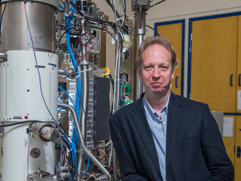
Angus Kirkland, JEOL Professor of Electron Microscopy and Secretary General of the International Federation of Societies for Microscopy, leads the Oxford Electron Image Analysis Group. His chief research interest is in the development of methods and instruments, including the theory and computation that accompany experiments, and their application to a range of materials problems.
Working with partners from the University, industry and overseas, Professor Kirkland is currently involved with three main areas of research: low-dimensional materials such as graphene, with its applications for structural materials and electronic device construction; nanoparticles, mainly relating to catalysts; and glass-like, amorphous materials.
These amorphous glasses, explains Professor Kirkland, ‘have potentially interesting bioactivity. There’s a lot of industrial interest at the moment in, for example, amorphous pharmaceuticals – non-crystalline versions of drug formulations.
‘They are not crystalline, ordered arrays of atoms; they are essentially a quasi-random array of atoms in the material. So they are very difficult to understand structurally. One problem is that they are incredibly radiation sensitive, so they decompose very rapidly under the electron beam.’
Professor of Materials Peter Nellist, President of the Royal Microscopical Society, shares Professor Kirkland’s interest in catalysis. ‘Catalysts’, he notes, ‘are incredibly important. They are responsible for the vast majority of industrial chemical processes in the world.’ They are used in the automotive sector (in car exhaust systems, for example) and the petrochemical sector, for refining. They also are required for hydrogen fuel cells – a promising technology for controlling pollution.
Professor Nellist explains: ‘A lot of catalysts consist of nanoparticles. To understand how a catalyst works, you need to be able to see the atoms within the nanoparticles – and the only way to do that is through electron microscopy. That then enables us to make better catalysts.’
Professor Kirkland adds that full characterisation of many catalysts requires imaging under gas environments, being developed as part of the collaboration with JEOL.
Professor Nellist’s group is also investigating the development of new lighting technologies. Lighting currently uses almost 20% of the world’s energy production. The latest research focuses on light-emitting diode (LED) technology, based on the substance gallium nitride.
Not naturally occurring, gallium nitride has to be grown in a laboratory – but the crystals have a very high incidence of defects, known as dislocations. Although the gallium nitride is still usable, the dislocations reduce the efficiency and lifetime of the LEDs.
‘We’ve been seeking to understand’, says Professor Nellist, ‘exactly how the atoms are arranged in these defects, and then how we can control them and try to reduce how they affect the lighting.’
In the last 15 years we’ve seen a real quantum leap in the capabilities of instruments.Professor Peter Nellist
The relationship with JEOL inspires both sides, with JEOL developing technology to support new research ideas.
One example of this is a new type of camera for microscopes, able to take up to an incredible 20,000 pictures a second. The technology was suggested by JEOL for a ptychography technique Professor Nellist was interested in, which requires a very fast camera in order to take a large four-dimensional data set from the microscope.
Using the new camera, the technique Professor Nellist’s group has now developed enables researchers to look at a wider range of different atom types within a sample.
He points out that ‘carbon atoms in particular can be quite hard to detect, because carbon is a very light element – it doesn’t scatter electrons very strongly. So this has really broadened the range of materials that the microscope can be used to look at.’
The camera has recently enabled research into complicated carbon nanotubes in collaboration with the Department of Chemistry.
A second important development in the field, again developed with JEOL, is the electron monochromator. This provides a low-energy spread in the electron beam, resulting in clearer images. It has been used to record images of individual defects in graphene.
This type of support enables us to do those adventurous kinds of research that would be harder to fund through conventional routes.Professor Peter Nellist
Professor Kirkland stresses that the flexible nature of JEOL’s support ‘has been incredibly useful for us because it’s not tied to a particular research theme; it’s not ringfenced to any particular spending profile. It’s very flexible, very responsive funding. So we don’t have to second-guess at the start about what we’re going to need for the next five years. We can do that as we go along – which is hugely important.’
Professor Nellist concludes: ‘This type of support enables us to do those adventurous kinds of research that would be harder to fund through conventional routes.’